Ultrasound
Need an Ultrasound in Hudson County, NJ?
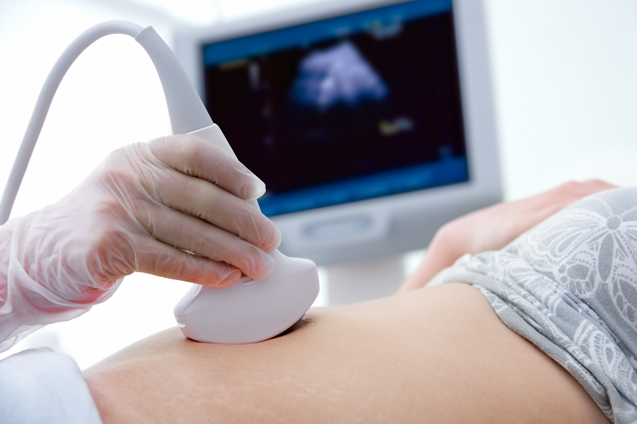
Ultrasound machines, also known as ultrasound scanners or sonography machines, are medical devices that use high-frequency sound waves to create images of the inside of the body. These machines consist of a transducer, which emits the sound waves, and a computer system that processes the returning echoes to generate images in real-time.
Ultrasound imaging is non-invasive and does not use ionizing radiation, making it a safe and widely used tool in medical diagnostics. It can be used to visualize various organs and tissues throughout the body, including the abdomen, pelvis, heart, blood vessels, muscles, tendons, and joints. In obstetrics and gynecology, ultrasound is commonly used to monitor fetal development during pregnancy.
Ultrasound machines come in various sizes and configurations, from portable handheld devices to larger console-based systems. They offer versatility in imaging, allowing healthcare professionals to perform diagnostic evaluations, guide minimally invasive procedures such as biopsies or injections, and monitor treatment progress.
Overall, ultrasound machines play a crucial role in medical practice by providing real-time imaging for diagnostic purposes, guiding interventions, and monitoring patient health without the need for ionizing radiation or invasive procedures.
Address
244 St Pauls Ave, Jersey City, NJ 07306
Call Us
(201) 201-3500
Email Us
office@njmri.com
Understanding Ultrasounds: A Patient Guide
An ultrasound is a non-invasive imaging technique that uses high-frequency sound waves to produce images of the inside of your body. It helps doctors diagnose and monitor medical conditions without the use of radiation. Here’s what you need to know before your ultrasound appointment at our Radiology Clinic:
What is an Ultrasound?
Ultrasound imaging, also known as sonography, allows your doctor to see soft tissues, organs, and blood flow throughout the body. It’s commonly used during pregnancy but is also used for examining the heart, kidneys, liver, and other organs.
Preparing for Your Ultrasound
- Instructions: Depending on the area being examined, you might need to fast for several hours or have a full bladder. Your clinic will provide specific instructions.
- Clothing: Wear comfortable clothing that allows easy access to the area being examined. You may be asked to change into a gown.
- Personal Items: You should remove any jewelry or other items that might interfere with the ultrasound.
During the Scan
- Positioning: You’ll lie on an examination table, and a technician (sonographer) will apply a special gel to the area being examined. This gel helps conduct sound waves.
- Procedure: The sonographer moves a device called a transducer over your skin. The transducer emits sound waves and captures the echoes that bounce back, creating images on a monitor.
- Time: The procedure typically takes 30 to 60 minutes, depending on the area being examined.
After the Scan
- Results: A radiologist will analyze the ultrasound images and send a report to your doctor, who will discuss the findings with you.
- Routine: You can return to your normal activities immediately after an ultrasound.
Safety and Comfort
Ultrasounds are safe and painless. There’s no radiation exposure, making it a preferred method for examining developing fetuses in pregnant women. The procedure should cause no discomfort, though you may feel slight pressure as the transducer is moved over your body.
Our team at the Radiology Clinic is here to ensure your ultrasound experience is as comfortable and informative as possible. If you have any questions or concerns about your upcoming ultrasound, please feel free to contact us.
