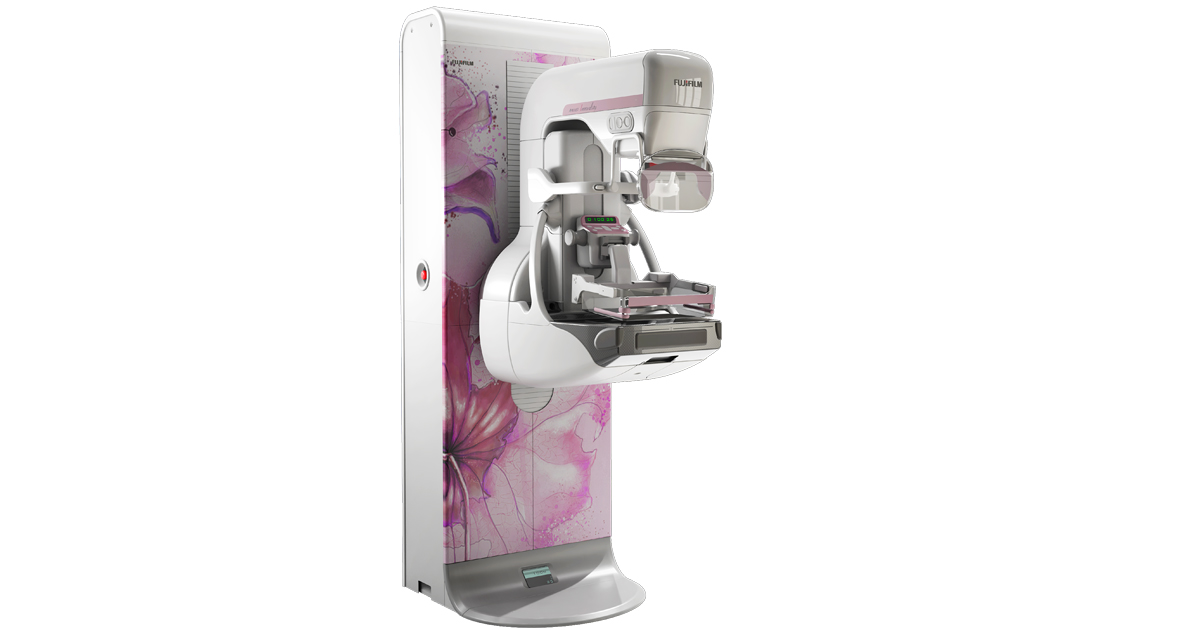
3D Mammogram at Apex Radiology
Need a Mammogram in Hudson County, NJ?
A 3D tomo mammography machine, also known as digital breast tomosynthesis (DBT), is an advanced imaging system used for breast cancer screening and diagnosis. Unlike traditional 2D mammography, which captures images of the breast in a single plane, 3D tomo mammography creates a series of thin, high-resolution images of the breast tissue from multiple angles.
This allows radiologists to examine the breast tissue layer by layer, reducing the overlap of structures that can obscure abnormalities and improving the detection of breast cancer. By providing a clearer view of breast tissue, 3D tomo mammography helps reduce false positives and false negatives, leading to more accurate diagnoses and potentially earlier detection of breast cancer. Overall, this technology represents a significant advancement in breast imaging, enhancing the effectiveness of breast cancer screening programs and improving patient outcomes.
Address
244 St Pauls Ave, Jersey City, NJ 07306
Call Us
(201) 201-3500
Email Us
office@njmri.com
Understanding Mammograms: A Patient Guide
A mammogram is a specialized X-ray of the breast used to detect and diagnose breast abnormalities, including cancer. It’s a crucial tool in breast cancer screening and can help detect tumors that cannot be felt and other changes in the breast.
Before Your Appointment
- Preparation: Avoid using deodorants, antiperspirants, powders, lotions, or perfumes under your arms or on your breasts on the day of the mammogram, as these can appear as white spots on the X-ray.
- What to Wear: Dress comfortably, preferably in a two-piece outfit, since you will be asked to undress from the waist up and wear a gown provided by the clinic.
- Scheduling: If you still menstruate, you might want to schedule your mammogram for the week after your period when your breasts are less tender, making the exam less uncomfortable.
During the Procedure
- Procedure: At the clinic, you’ll stand in front of a mammography machine, and a technician will place your breast on a clear plastic plate. Another plate will firmly press your breast from above. The plates flatten the breast, spreading the tissue apart to get a clearer image. The process is repeated to take images from multiple angles. You’ll feel pressure on your breast for a few seconds, which can be uncomfortable or slightly painful for some.
- Duration: The entire process usually takes about 20 to 30 minutes, with the actual breast compression lasting just a few seconds per image.
After the Procedure
- Results: A radiologist will analyze the mammogram images and send a report to your doctor, who will discuss the results with you. If there’s anything unusual, you may need additional imaging tests.
- Follow-Up: Depending on your results, your doctor may recommend a routine follow-up mammogram in one to two years or further diagnostic tests if any abnormalities are found.
Additional Information
- Safety: Mammograms use a low dose of radiation to create images of the breast. The risk from this radiation exposure is considered very small compared to the benefits of detection and prevention of breast cancer.
- Screening Recommendations: Guidelines vary, but it’s commonly recommended for women to begin screening mammograms at age 40 and continue annually or every two years through at least age 74, depending on individual risk factors and health.
It’s normal to feel anxious about getting a mammogram, but remember that it’s a crucial step in maintaining your breast health. If you have any concerns or questions, do not hesitate to discuss them with your healthcare provider. They can provide you with more information tailored to your specific health needs.
